A 60-year-old woman with a history of severe chronic thromboembolic hypertension presented with progressive symptoms of typical angina. Cardiac computed tomography angiography (CTA) revealed a filiform-shaped left main (LM) with severe systolic/diastolic vessel compression between a prominently enlarged pulmonary artery trunk (Ø68 mm) and the aortic sinus (Figure 1A-Figure 1C, Moving image 1, Moving image 2). Stress echocardiography showed an inducible ischaemia in the anterior wall. Invasive coronary angiography and intravascular ultrasound (IVUS) confirmed these results with a pulsatile 80% stenosis of the LM (Figure 1D). IVUS excluded coronary plaque stenosis (Figure 1E). A 5.0×12 mm Dynamic Renal stent (Biotronik, Berlin, Germany) used for its increased radial stability was placed in the LM. Post-interventional 4D-cardiac CTA showed good stent positioning and efficient vessel bridging during dynamic systolic/diastolic compression (Figure 1F-Figure 1I, Moving image 3). Post-interventional IVUS was used to confirm full stent expansion and wall apposition (Figure 1J). The patient was discharged in a good condition.
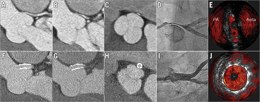
Figure 1. Assessment and intervention of left main compression by an enlarged pulmonary artery. Cardiac CTA showing the left main travelling in-between the aortic sinus and a large pulmonary artery (PA) with pulsatile systolic (A)/diastolic (B, C) spindle-shaped compression by the pulmonary artery and the left coronary cusp. Corresponding coronary angiography (D) and intravascular ultrasound (IVUS) (E) confirmed compression of the left main. Placement of a Dynamic Renal stent in the left main exhibited strong left main shielding during systole (F) and diastole (G, H) as shown with CTA and coronary angiography (I). IVUS confirmed full stent expansion and proper wall apposition (J).
Conflict of interest statement
The authors have no conflicts of interest to declare.
Supplementary data
Moving image 1. 4D-CTA of left main compression (cross-sectional view).
Moving image 2. 4D-CTA of left main compression (longitudinal view). 4D-cardiac CTA shows pulsatile systolic/diastolic compression of left main by an enlarged pulmonary artery.
Moving image 3. CTA of LM bridging after stenting. 4D-cardiac CTA of the left main after stenting shows vessel bridging during systolic/diastolic movement.
Supplementary data
To read the full content of this article, please download the PDF.
Moving image 1. 4D-CTA of left main compression (cross-sectional view).
Moving image 2. 4D-CTA of left main compression (longitudinal view). 4D-cardiac CTA shows pulsatile systolic/diastolic compression of left main by an enlarged pulmonary artery.
Moving image 3. CTA of LM bridging after stenting. 4D-cardiac CTA of the left main after stenting shows vessel bridging during systolic/diastolic movement.