Cory:
Unlock Your AI Assistant Now!
ST-segment elevation myocardial infarction (STEMI) with a large thrombus burden (LTB) remains a prognostically relevant clinical challenge. However, thrombus aspiration, the prevailing approach to thrombus modification during STEMI, has not shown efficacy in randomised studies. Meanwhile, in stroke intervention, stent retriever-assisted thrombectomy has demonstrated clear efficacy1.
Whether stent retriever-assisted thrombectomy could play a role in STEMI patients with an LTB remains unanswered. Only case reports suggest the feasibility of this approach, while ongoing studies aim to evaluate its effectiveness in enhancing thrombus extraction2.
Here we present unique images visualising the impact of stent retriever-assisted thrombectomy in a patient admitted with STEMI and LTB, and in a coronary bench model.
A 69-year-old male presented to our institution with an inferior STEMI due to a thrombotic occlusion of the left circumflex artery. Coronary flow was restored with a 2 mm semicompliant balloon, revealing a large filling defect at the site of occlusion on angiogram (Figure 1A, Moving image 1); this filling defect was confirmed to be a large volume of thrombus by optical frequency domain imaging (OFDI; Lunawave [Terumo]) (Figure 1B, Moving image 2). A stent retriever was successfully deployed (Figure 1C), following the procedural steps described by Spirito et al3. A large red thrombus was extracted (Figure 1D), and subsequent angiography and OFDI demonstrated full clearance of thrombus at the site of the lesion (Figure 1E, Figure 1F, Moving image 3, Moving image 4). Finally, a 3.5x20 mm drug-eluting stent was deployed and postdilated with a 3.5 mm non-compliant balloon.
To dissect the mechanism of action of the stent retriever, we used a coronary bench model, using a cylinder of spreadable butter to simulate a fully occlusive thrombus. We employed intravascular imaging (OFDI) to assess the impact of stent retriever-assisted thrombectomy and manual thrombus aspiration on the simulated thrombus (Figure 1G, Figure 1H). Moving image 5 shows the deployment of a stent retriever device (Solitaire X [Medtronic]) in the coronary model with extraction of the simulated thrombus. Moving image 6 replicates the same bench model, with manual thrombus aspiration (Export Advance [Medtronic]).
OFDI following stent retriever-assisted thrombectomy showed near-complete extraction of the simulated thrombus, with detection of stent retriever strut-shaped indentations embedded in the residual simulated thrombus and abutting the vessel wall circumferentially, suggesting that the device had expanded to the size of the vessel (Figure 1G). These indentations were not seen in the clinical case (Figure 1F) due to the differing consistency of a real thrombus. OFDI performed in the bench model following manual thrombus aspiration demonstrated that a core of simulated thrombus had been extracted but a large volume of residual material had been left behind (Figure 1H).
The use of stent retriever devices for thrombus modification in STEMI with LTB is feasible. Results from clinical trials systematically evaluating this interventional strategy are awaited.
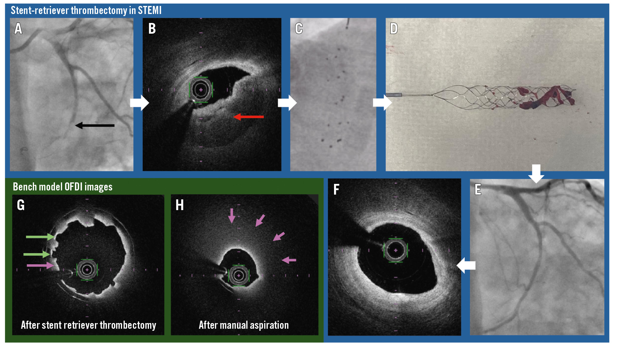
Figure 1. Stent retriever-assisted thrombectomy in STEMI. A) Angiogram showing a filling defect (indicated by the black arrow) in the left circumflex in a patient presenting with inferior STEMI. B) OFDI frame prior to thrombectomy showing a large coronary thrombus burden (indicated by the red arrow). C) The expanded stent retriever in the left circumflex. D) Extracted red coronary thrombus attached to stent retriever. E) Angiogram after stent retriever-assisted thrombectomy. F) OFDI frame showing the same segment of vessel as B, after stent retriever-assisted thrombectomy, with no thrombus visible. G) OFDI image after stent retriever-assisted thrombectomy in a coronary model. Green arrows indicate stent retriever indentations remaining in the simulated thrombus (butter). The purple arrow indicates residual thrombus. H) OFDI image after aspiration in a coronary model. Purple arrows indicate the large volume of residual simulated thrombus. OFDI: optical frequency domain imaging; STEMI: ST-segment elevation myocardial infarction
Acknowledgements
We would like to acknowledge Professor Keith Channon, who was the operator for the case presented, and Professor Adrian Banning for his helpful comments during the preparation of this manuscript.
Conflict of interest statement
All the authors have received a grant or research support (to their institution) from Medtronic and Terumo.
Supplementary data
To read the full content of this article, please download the PDF.
Moving image 1. Angiogram showing a filling defect and slow flow in the circumflex artery.
Moving image 2. OFDI run of the circumflex artery prior to thrombectomy.
Moving image 3. Angiogram showing resolution of filling defect and improved flow in the circumflex artery.
Moving image 4. OFDI run of the circumflex artery after thrombectomy.
Moving image 5. Bench model of a coronary artery showing a stent retriever extracting a simulated thrombus.
Moving image 6. Bench model of a coronary artery showing an aspiration catheter extracting a simulated thrombus.
