DOI:

Multifocal arterial fibromuscular dysplasia causing coronary artery dissection following coronary angiography

Jonas de Jong*, MD; Jan Piek, MD, PhD; Allard van der Wal, MD, PhD

A 31 year old man was referred to our hospital for evaluation of recurrent angina pectoris following recent inferior-posterior myocardial infarction. His previous medical history included a surgically corrected tetralogy of Fallot at four years of age. During coronary angiography of the left anterior descending artery, a dissection of the entire vessel developed (Figure 1).
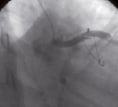
Figure 1 and Movie 1. Acute dissection of the LAD during diagnostic catheterisation.

The dissection could not be crossed with a guidewire and the patient died.

Post mortem examination was performed. Histologically, the LAD dissection channel was filled with fresh haematoma indicating recent onset (Figure 2).

Figure 2. Cross section of the LAD showing a dissection channel in the outer part of the arterial media, filled with haematoma (Mallory’s stain).

The media showed abnormal presence of segmental ridges of medial tissue containing haphazardly arranged smooth muscle layers intermingled with elastin and collagenous tissue (Figure 3).

Figure 3. Detail of the coronary artery wall showing highly irregular medial ridges of fibromuscular tissue characteristic for the medial type of FMD. (Elastica van Gieson stain).

There were no signs of of inflammation (Figures 2 and 3) or previous dissections. These findings were diagnostic for the media type of fibromuscular dysplasia (FMD).

FMD is a segmental non-atherosclerotic non-inflammatory angiopathy of unknown aetiology. Renal and cervicocranial arteries are most commonly involved. Coronary FMD is very rare1. The disease is histologically classified according to the principal layer of arterial involvement (intimal-, medial- and peri-adventitial FMD). Medial FMD as found in our patient is by far (approximately 90% of cases) the commonest type.

Reference
Volume 5 Number 1
May 19, 2009
Volume 5 Number 1
View full issue


Key metrics

Suggested by Cory

Editorial

10.4244/EIJ-E-24-00065 Apr 21, 2025
From invasive gradients to pressure recovery: rethinking long-standing paradigms
Joner M and Mylotte D
free

Original Research

10.4244/EIJ-D-24-00341 Apr 21, 2025
Prognostic value of invasive versus echocardiography-derived aortic gradient in patients undergoing TAVI
van den Dorpel M et al

Debate

10.4244/EIJ-E-24-00071 Apr 21, 2025
Could the age threshold for TAVI be relaxed to below 65 years? Pros and cons
Garot P et al

Editorial

10.4244/EIJ-E-25-00011 Apr 21, 2025
Transcatheter aortic valve implantation with complex, high-risk indicated PCI
Patterson T and McDonaugh B
free

State-of-the-Art

10.4244/EIJ-D-24-00066 Apr 21, 2025
Management of complications after valvular interventions
Bansal A et al
free
Trending articles
69.996

10.4244/EIJV13I12A217 Dec 8, 2017
Swimming against the tide: insights from the ORBITA trial
Al-Lamee R and Francis D
free
59.65

State-of-the-Art

10.4244/EIJ-D-24-00066 Apr 21, 2025
Management of complications after valvular interventions
Bansal A et al
free
57.6

State-of-the-Art

10.4244/EIJ-D-24-00386 Feb 3, 2025
Mechanical circulatory support for complex, high-risk percutaneous coronary intervention
Ferro E et al
free
38.75

State-of-the-Art

10.4244/EIJ-D-23-00912 Oct 7, 2024
Optical coherence tomography to guide percutaneous coronary intervention
Almajid F et al
free
15.85

State-of-the-Art

10.4244/EIJ-D-23-01050 Jul 15, 2024
Durability of transcatheter aortic valve implantation
Ternacle J et al
free
X

The Official Journal of EuroPCR and the European Association of Percutaneous Cardiovascular Interventions (EAPCI)

EuroPCR EAPCI
PCR ESC
Impact factor: 7.6
2023 Journal Citation Reports®
Science Edition (Clarivate Analytics, 2024)
Online ISSN 1969-6213 - Print ISSN 1774-024X
© 2005-2025 Europa Group - All rights reserved